Case Study of A Rare Case with Tessier Cleft, Hypertelorism and Bifid Nose
Recently, at Richardson’s Hospital, we encountered a rare case posing unique clinical features with multiple challenges. We are grateful that we had the privilege to perform such a life-transformative surgery on an 11-year-old girl.
She had already undergone surgery for ethmoidal encephalocele at another hospital before she came to us. Following her initial surgery for ethmoidal encephalocele, she faced lingering challenges with significant facial and nasal deformities.
Tessier Cleft, Hypertelorism and Bifid Nose
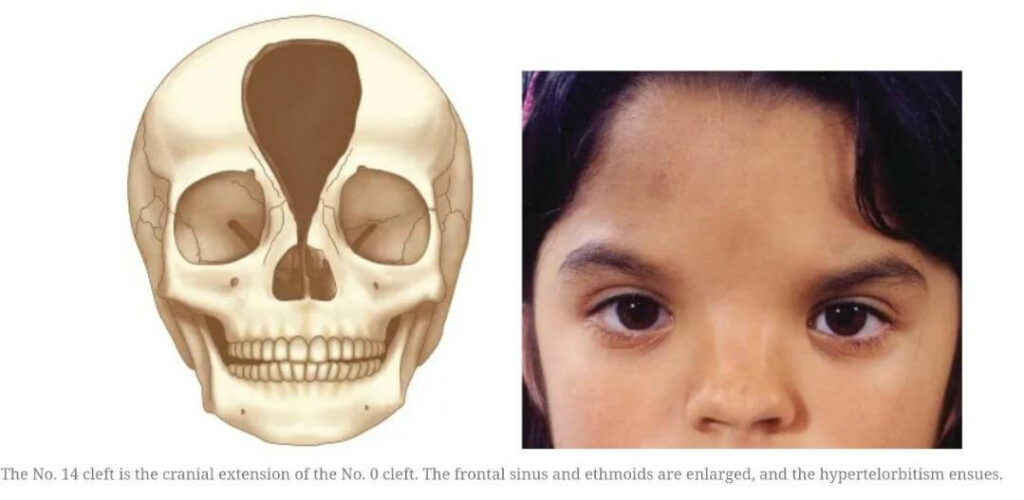
The patient presented a complex craniofacial scenario, marked by Tessier cleft no. 14 and a history of ethmoidal encephalocele surgery. The facial challenges were compounded by a bifid nose and hypertelorism, leading to vision abnormalities. Understanding the interplay of these factors was crucial in planning an effective intervention and addressing all these issues.
What Are Craniofacial Anomalies?
Craniofacial anomalies are conditions that affect the head and face, often present from birth. These anomalies involve irregularities in the development of bones and tissues in the skull and facial region. In simpler terms, they are variations from the typical appearance of a person’s head and face.
Craniofacial anomalies may impact both appearance and function. Some anomalies are mild and primarily cosmetic, while others can affect breathing, hearing, or even brain development.
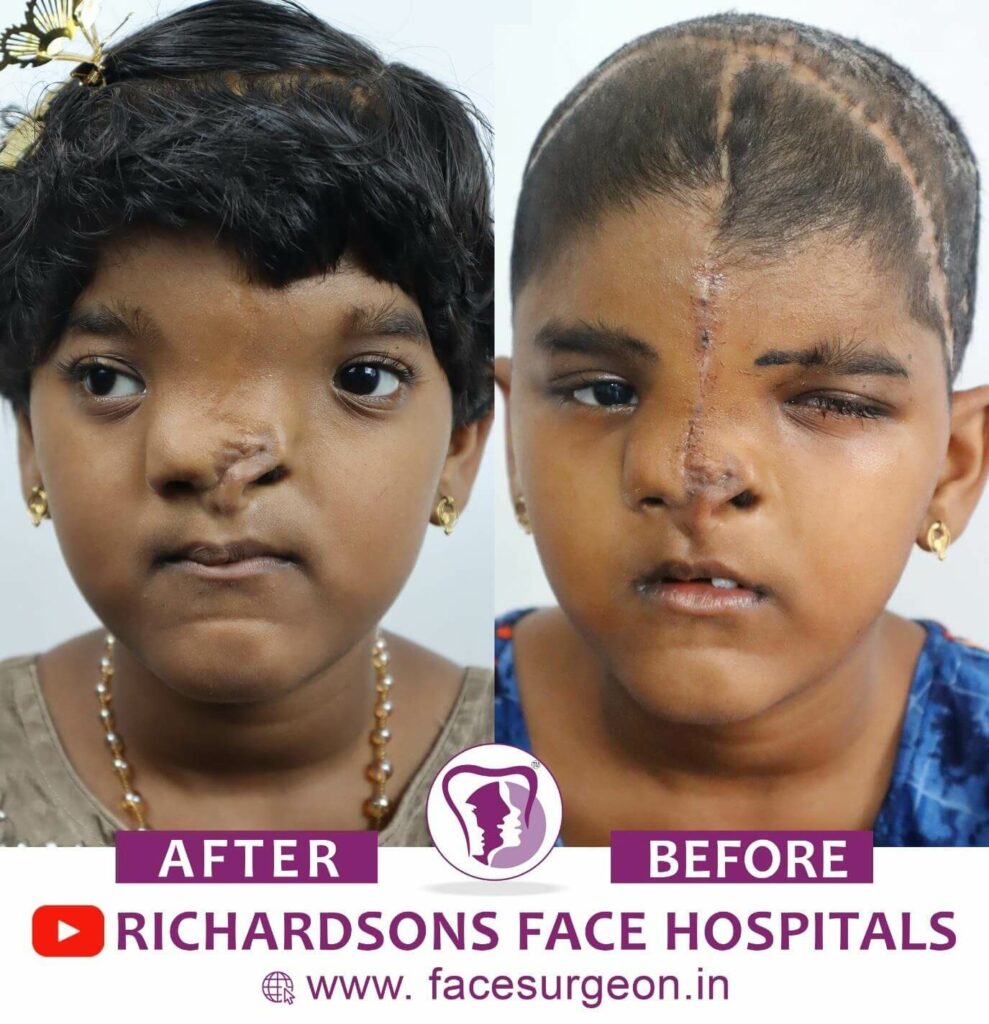
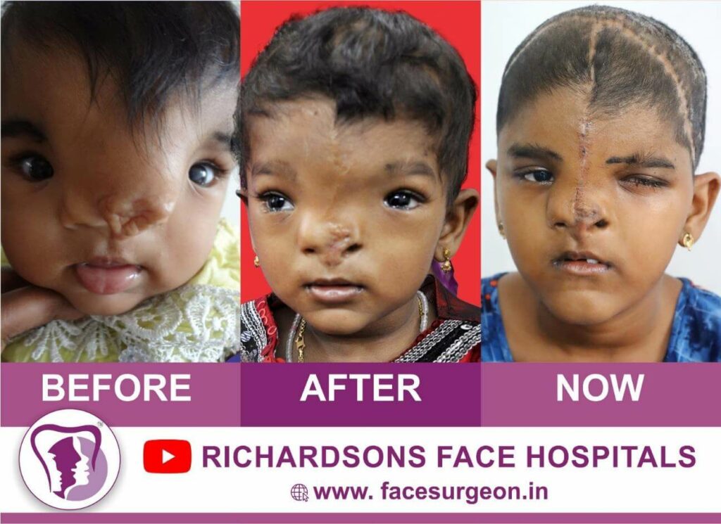
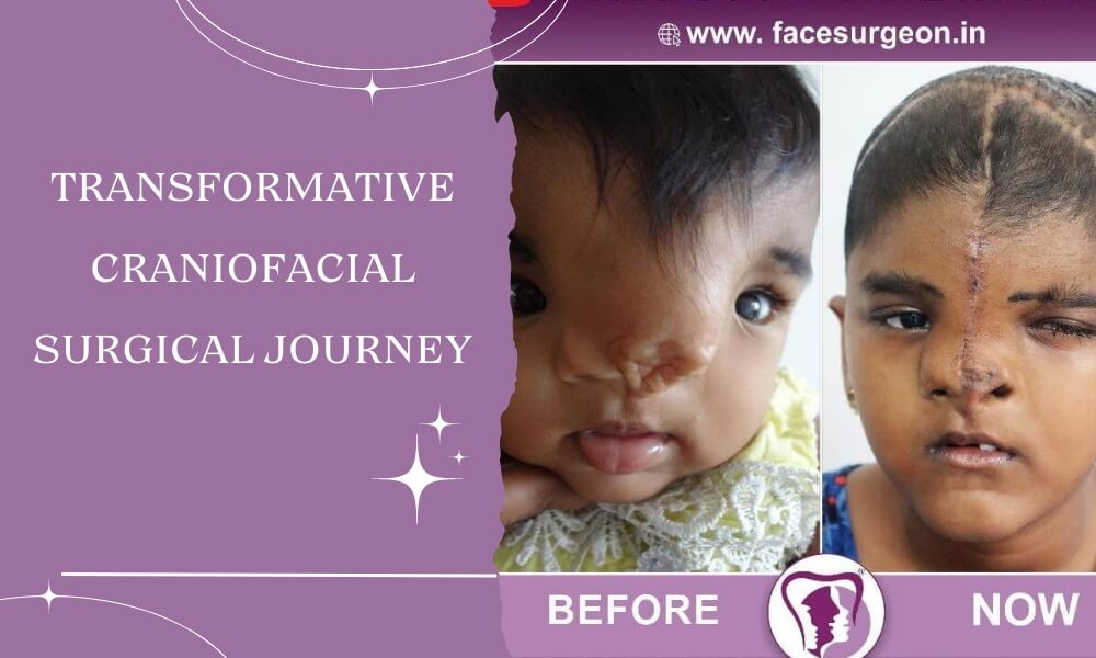
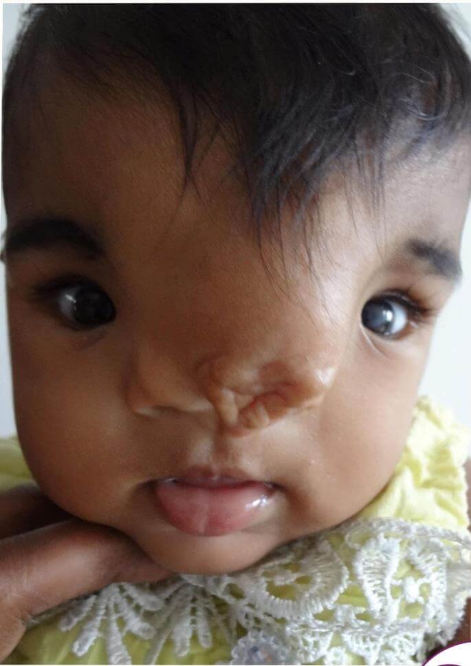 This is how she looked because of the craniofacial deformity she had since birth.
In the image above the following were the facial features which needed surgical intervention for correction:
- Hypertelorism.
- Tessier Cleft.
- Depressed, Broad, and flat forehead and glabella.
- Nasal soft tissue and Bone deformities.
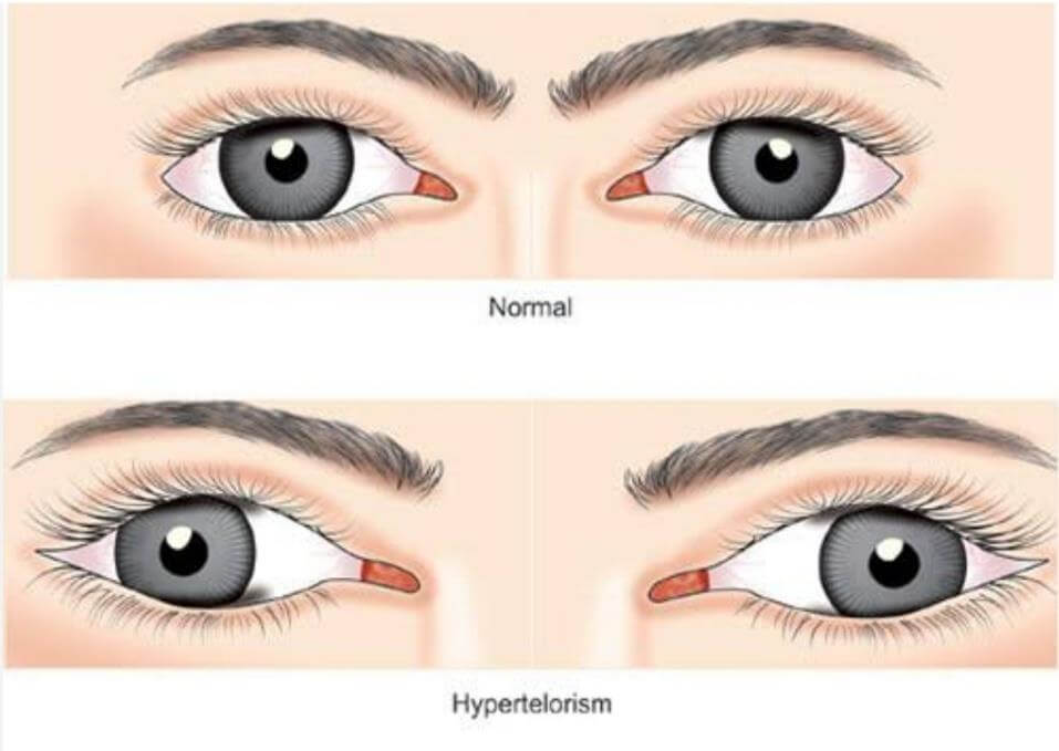
What is Hypertelorism?
It’s a condition where the space/gap between the eyes is larger than usual, and this can give a distinct appearance to the face. It is like your eyes are set wider apart on your face.
Instead of having a normal distance between their eyes, there’s an increased abnormal distance.
Tessier clefts are rare congenital facial clefts or cleft-like deformities that occur during fetal development. Tessier clefts can affect various facial structures, including the eyes, nose, and mouth.
Connection
The connection between hypertelorism and Tessier clefts lies in the fact that some Tessier clefts can involve the area around the eyes, leading to an increased distance between the eyes essentially, hypertelorism.
Surgical Strategy
Facial Bipartition Surgery
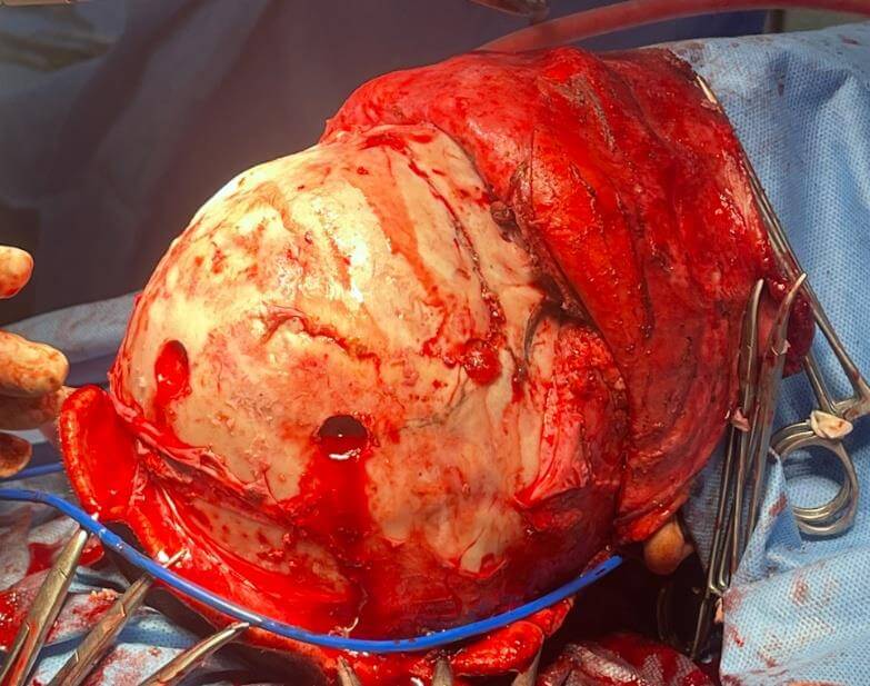
The cornerstone of the surgical intervention was a facial bipartition surgery, meticulously planned to correct the hypertelorism. The bi-coronal approach was chosen to ensure optimal visibility and access to the affected areas. This procedure involved carefully repositioning the facial and cranial bones to achieve symmetry and alleviate the patient’s vision abnormalities.
With the help of technological advancements, we had 3D printed the entire skull model of a patient which aided while doing surgical execution.
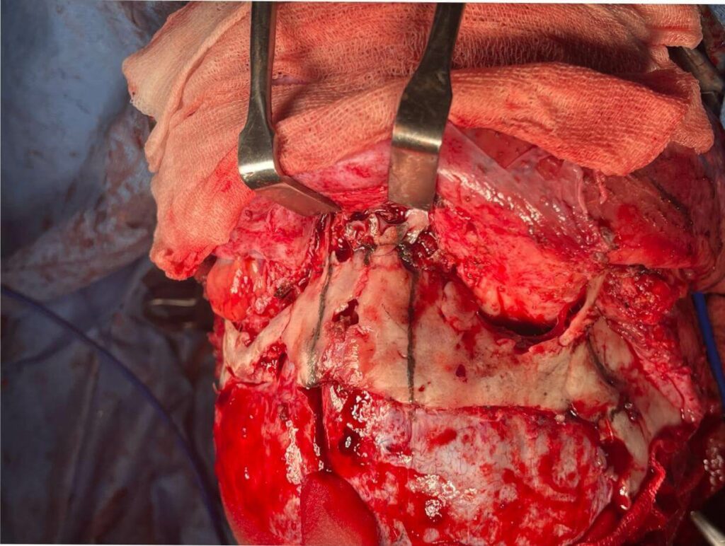
Facial bipartition surgery complex craniofacial procedure was done by performing controlled osteotomy, repositioning, and stabilization of the facial as well as cranial bones, particularly those constituting the midface and orbits.
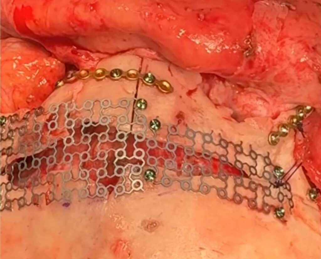
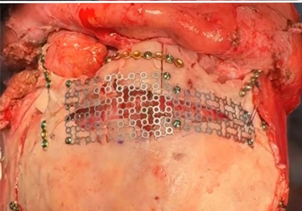
After exposing the underlying structures, osteotomies were strategically performed to separate the facial skeleton into two parts. This division allowed for to precise repositioning of the bony segments to normalize interorbital spacing and correct associated craniofacial abnormalities. Fixation of the mobilized craniofacial skeleton was done using titanium plates, screws, and mesh.
Also, a portion of the excess skin near the nasal bridge region was excised meticulously considering the aesthetic requirements.
Canthopexy
Addressing the hypertelorism also required a cathopexy, a surgical technique involving the repositioning/stabilizing of the medial canthus, which is the inner corner of the eye. The surgical techniques employed included the use of sutures for additional structural support for getting desired outcomes functionally and esthetically.
Post Operative Healing and Recovery
Following the surgery, the patient underwent a ten-day hospital stay for close monitoring and postoperative care. The recovery period was uneventful, reflecting the success of the surgical intervention and the comprehensive approach to patient management.
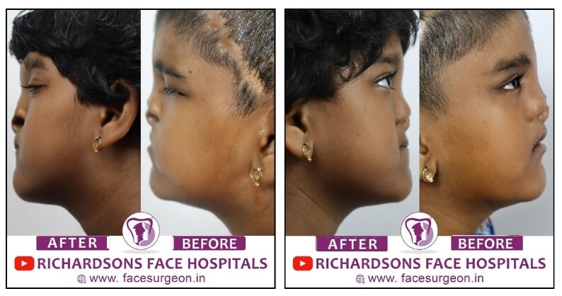
The outcomes of the facial bipartition surgery were nothing short of remarkable. The correction of hypertelorism not only restored facial symmetry but also addressed the vision abnormalities associated with the condition. The patient experienced a significant improvement in both form and function, highlighting the success of the surgical intervention. The depressed and flat forehead now got adequate projection and form.
Need for Subsequent Surgeries
As our patient progresses through her twenties and thirties, we anticipate the need for further refinement. In the future, a rhinoplasty / Nose surgery may be recommended to address any residual nasal deformities, ensuring a comprehensive and enduring solution for our patient’s evolving needs.
Our patient’s journey exemplifies the intricate nature of craniofacial surgery, where each phase is a building block in the pursuit of optimal form and function. Looking forward, we remain committed to providing comprehensive care for her.
